About ZALKAL#
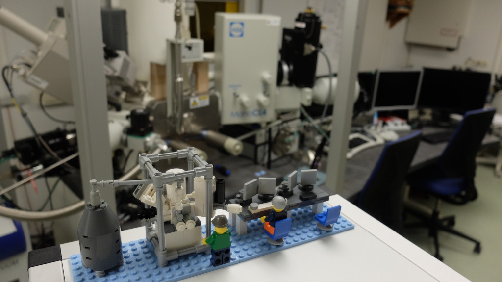
Application lab#
The Paul Drude Institute has 30 years of experience in the field of cathodoluminescence (CL) spectroscopy, which analyzes the light emitted by semiconductors under electron irradiation. The light is emitted when charge carriers (electrons and holes) recombine and its wavelength is determined, among other things, by the band gap of the semiconductor. Based on a scanning electron microscope (SEM), this offers the possibility to investigate properties of semiconductor structures with extremely high spatial and spectral resolution. On the one hand, this allows a more detailed understanding of structural defects in the crystal structure as they influence the light emission and thus their influence on desired properties can be investigated. On the other hand, nanoscopic structures can be investigated that have been incorporated into the semiconductor to tailor its functionality. However, knowledge on the dynamics, i.e. the temporal behavior of the charge carriers, which cannot be investigated with a classical CL system, are crucial for a comprehensive understanding of where charge carriers are lost, i.e. efficiency is lost in the structures. The required high temporal resolution is achieved by pulsing the electron beam on a picosecond timescale combined with ultrafast detectors.
The PDI is currently building up an application laboratory for time-resolved cathodoluminescence spectroscopy. The aim is to provide the best possible spatial, spectral and temporal resolution, where the chosen detectors will be optimized for ultraviolet wavelengths down to about 180 nm. However, the visible and near-infrared spectral regions will be covered as well, thus covering a wide range of materials. The system will be capable of measuring classic semiconductor thin-films and heterostructures, 2D materials, as well as 3D nanostructures. It thus can contribute to a wide range of the research activities at PDI. However, we will also closely collaborate with other research institutes and academic partners working on semiconductors in Berlin and beyond, as well as companies that require the combination of spectral and time-resolved luminescence mapping. Using the existing infrastructure of the analytical SEM lab, measurements can be correlated to maps of the composition (energy-dispersive x-ray spectroscopy, EDX), crystal structure (electron backscatter detection, EBSD) and charge collection (electron beam induced current, EBIC).
The application lab is co-funded by the European Regional Development Fund through the State of Berlin (Senate) from January 2023 through December 2025.
Fundamentals#
Semiconductors#
The band structure in a crystal originates from the overlap of atomic orbitals when many atoms are brought together to form a solid. As atoms approach each other, their outer (valence) orbitals interact and split into a very large number of closely spaced energy levels, forming continuous energy bands rather than discrete atomic levels. In a semiconductor, a special case arises: a lower band (valence band), formed from filled atomic orbitals, is completely occupied by electrons, while the next higher band (conduction band) is empty at low temperature. The finite energy gap between the highest filled state and the lowest empty state is called the bandgap. This bandgap prevents electrons from conducting electricity unless they are excited across this gap by the supply of energy (e.g. thermal, optical, electrical excitation).

Origin of the bandgap of a semiconductor from the overlap of atomic levels in a crystal leading to energy bands of which one is completely filled
Scanning electron microscopy#
In a scanning electron microscope (SEM), the surface of a sample is scanned with a focused electron beam. The low-energy secondary electrons excited near the sample surface are measured by a detector and thus the sample surface is imaged. Secondary electrons excited deeper in the sample are reabsorbed before they can leave the sample. This allows structures to be visualized that are not visible in a light microscope due to the diffraction limit.
The interaction of the incident high-energy electrons with a solid produces a whole cascade of scattering processes. In addition to secondary electrons some of the energy is for example emitted in the form of X-rays, which enables additional measurements in electron microscopy. Among other things, the characteristic X-rays make it possible to analyze the sample composition. The extent of the interaction volume in which these scattering processes take place depends on the energy of the incident electrons: the higher the energy (acceleration voltage, usually a few keV), the deeper and wider the volume in which the energy transfer takes place. As a consequence, the spatial resolution of these additional signals is lower than with surface imaging.

Schematic depiction of a scanning electron microscope

Interaction volume and signals in scanning electron microscopy
Cathodoluminescence spectroscopy#
Semiconductors are also excited by the electron beam to emit characteristic light, known as cathodoluminescence (CL). The decisive factor here is that semiconductors have a band gap: If enough energy is supplied to the electrons bound in the crystal to overcome this band gap, the semiconductor becomes conductive. This excitation process is at the end of the excitation cascade in an electron microscope, as only a few eV are required with the energy scaling with the band gap. Therefore, a single irradiated electron can excite several hundred conduction electrons. If such a conduction electron returns to a bound state in the so called valence band, the corresponding energy is released. This recombination can take place radiatively by emitting a photon (light particle) or non-radiatively when the energy is converted into heat (lattice vibrations). Depending on the semiconductor, the energy of the band gap corresponds to infrared (low energies), visible or ultraviolet (high energies) light.

CL excitation and recombination process in a semiconductor of band gap Eg

Band gap energies of some common semiconductors and correspondence to the light spectrum
On the one hand, the wavelength (color) of the light is also influenced by built-in foreign atoms (doping) or crystal defects. These introduce additional states in the bandgap and facilitate emission at lower energies (longer wavelengths). On the other hand, the composition of certain semiconductor structures (e.g. layers for light-emitting diodes) is varied to adjust the color of the light. Here, the different bandgap of such a heterostructure changes the emission energy. The spectral fingerprint therefore allows a variety of conclusions to be drawn about the quality and properties of semiconductor structures. If this light is collected and analyzed in a spectrometer, intensity changes and changes in wavelength can be locally resolved and imaged using an appropriately equipped electron microscope.


Missing or dopant atoms in the crystal structure introduce additional states in the bandgap


Heterostructure with different composition locally modifying the bandgap energy

Schematic representation of the main components of a CL-setup
The special feature of cathodoluminescence spectroscopy is the high spatial resolution of a few nanometers, which is achieved in a scanning electron microscope. Although the resolution of cathodoluminescence spectroscopy is limited by the above-mentioned generation volume and the additional diffusion of charge carriers in the semiconductor, it still exceeds the local resolution of photoluminescence spectroscopy by 1-2 orders of magnitude, depending on the material and measurement conditions. At the same time, a directly correlated image of the surface structure is always obtained.
In accordance with the functioning principle of electron microscopes, hyperspectral CL mapping involves moving the electron beam point by point across the sample and recording a complete luminescence spectrum at each grid point.

Animation of hyperspectral CL mapping: The electron beam scans the sample and a CL spectrum is collected at every position
Time-resolved spectroscopy#
The temporal decay of a luminescence signal after a short (pulsed) excitation contains important complementary information about the recombination processes and the properties of the semiconductor. This process can take place in a few ps or extend over many ns. In addition to this lifetime (time scale), the shape of the decaying intensity function and whether there are spectral shifts over time is also informative. Among other things, this can help to understand efficiency losses in semiconductor components.
Time-resolved excitation#
There are two ways to achieve pulsed excitation in an electron microscope. In the ZALKAL application laboratory, both options are implemented due to their complementary properties. The pulse duration must be shorter than the lifetime to be measured:

Comparison of standard SEM operation with the two complementary pulsing schemes
Electromagnetic deflection of the electron beam (ultrafast beam blanking). By correctly placing the deflection coils and using an aperture to cut off the deflected beam (so that it does not travel over the sample), pulse durations of less than 50 ps can be achieved. Since the continuous beam is manipulated here, this mode is very stable over long times, but the available beam current for which the short times are achieved is limited.
Laser-initiated pulsing of the electron source (cathode). If the cathode is operated below the emission threshold, an electron pulse can be triggered with a pulsed UV laser beam. With appropriate lasers, pulse durations of a few ps (targeted by ZALKAL) or even several hundred fs can be achieved. However, the laser must be precisely aligned with the cathode and kept stable over time.
Time-resolved detection#
The two standard methods for time-resolved detection of luminescence are time-correlated single photon counting (TCSPC) and the use of streak cameras. Both methods allow the measurement of temporal changes in luminescence intensity on time scales in the pico- or nanosecond range. In cathodoluminescence spectroscopy, in addition to direct detection of the decay dynamics, the autocorrelation function can also be measured to measure lifetimes.

Schematic comparison of the different time-resolved detection modes in cathodoluminescence spectroscopy
The autocorrelation function describes the temporal correlation of the emitted photons – that is, it indicates how likely it is to detect photons generated by the same excitation event at two different points in time. Since in cathodoluminescence a single electron can excite several hundred electron-hole pairs and thus photons, the emission time of these photons is correlated. The delay statistics between these correlated photons represent the lifetime of the recombination process. What is special here is that time-resolved measurements are possible without pulsed excitation. At the same time, however, the method is not suitable for particularly short or long lifetimes and only achieves limited dynamics (the orders of magnitude over which the decay curve can be measured). It also requires dedicated mathematical modeling of the correlation function.
Time-correlated single photon counting measures the arrival times of individual photons after a periodic excitation. Statistically, less than one photon is registered per excitation cycle, so that the probability of double registrations can be neglected. This results in a histogram of arrival times, which represents the lifetime of the luminescence. The temporal resolution is limited by the detectors (fast photomultipliers) and is therefore in the range of 20-200 ps. This detection method is particularly suitable for low signal intensities or to achieve high temporal dynamics. It is also the most suitable method to achieve lifetime mapping with shorter measurement times (with limited dynamics).
Streak cameras use an electron-optical arrangement to convert the temporal course of an incoming light signal into spatial information. The light signal is first converted into an electron beam, which can then be deflected by means of a time-variable electric field, and then converted back into a light signal with the aid of a photocathode, which is finally detected by a CCD camera. If the spectral splitting of the light by a spectrometer is used on the second axis, this directly results in a two-dimensional image of the luminescence of wavelength versus time. Streak cameras also enable extremely high time resolutions of a few picoseconds. However, splitting the light into two dimensions requires longer measurement times and results in lower dynamics. It also takes a larger effort to adjust the light incoupling.

Spectrally and temporally resolved image of the luminescence recorded with a streak camera
Team#
Project leader: | Dr. Jonas Lähnemann
Postdocs: | Dr. Kagiso Loeto (since 12/2023) | Dr. Mikel Gomez (since 03/2025)
PhD students: | Aiden Campbell (since 05/2024) | Domenik Spallek (since 06/2023) | Mikel Gomez (09/2020-03/2025)
Master students/Interns/Student assistants: | Joel Maillet (since 09/2025) | Daniel Ramesh Paulo-Wach (05/2025-09/2025) | Jonathan Valenzuela (09/2024-12/2024) | Aiden Campbell (09/2023-04/2024) | Johannes Pietsch (06/2022-10/2023)
Project administration: | Anja Holldack
