Facilities#
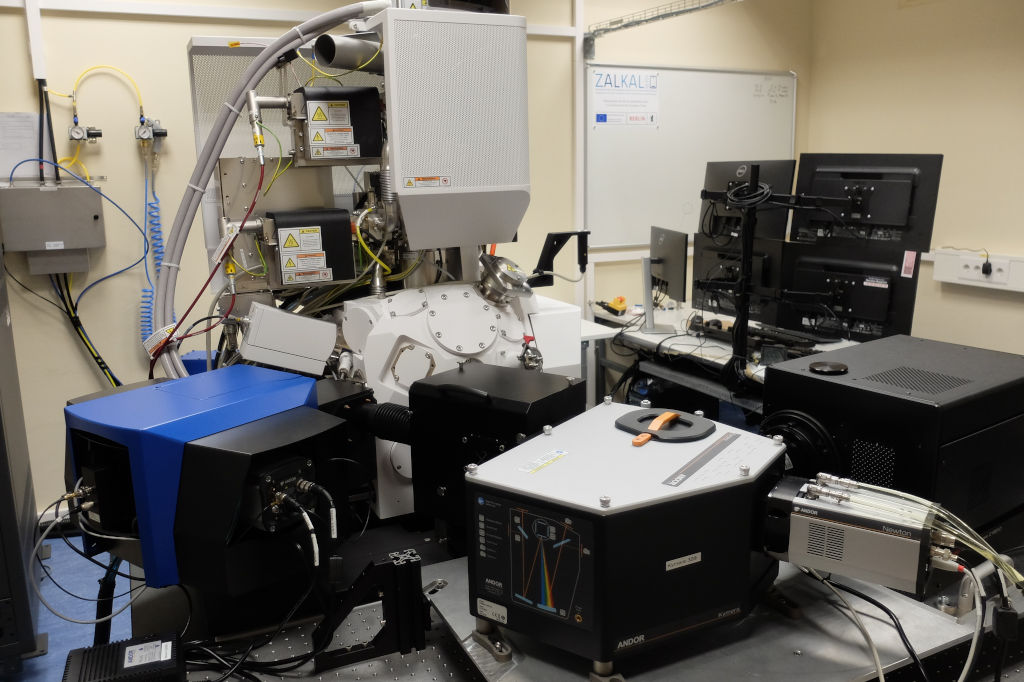
In the context of the application laboratory ZALKAL a dedicated scanning electron microscope for time-resolved cathodoluminescence spectroscopy (TRCL) was installed at Paul Drude Institute in 2024/2025. In addition, the PDI operates another analytical scanning electron microscope to correlate further spatially-resolved methods with the CL/TRCL measurements.
Time-resolved cathodoluminescence spectroscopy#
UV-optimized cathodoluminescence (CL) spectroscopy system with time-integrated and time-resolved detection based on a scanning electron microscope (SEM) with pulsed electron beam and helium-cooled sample stage for measuring hyperspectral maps, as well as the lifetime of charge carriers in semiconductor materials, hetero- and nanostructures in the spectral range of 180-1050 nm.
Main specifications#
Thermo Fisher Verios 5 UC field emission scanning electron microscope
Excitation
Acceleration voltage: 350 V to 30 kV
Beam current (continuous): 0.8 pA to 100 nA
Beam diameter <1 nm
Ultrafast beam blanking: <30 ps
Laser-pulsed cathode (2 ps) planned
Other characteristics
He cold stage (10–300 K)
Electron beam-induced current (EBIC) measurements with triax-cabeling
Electron detectors: Everhart-Thornley, Through-lens, In-Column and Transmission
In-Situ plasma-cleaner
Delmic Sparc Spectral CL system extended by second spectrometer and time-resolved detection
UV/vis-spectrometer 180–850 nm
328 mm focal length
CCD with quantum efficiency > 40% from 200–800 nm (Andor Newton 940 BU2)
Hamamatsu streak camera with MgF₂ photocathode 180–830 nm, 800 fs time resolution
If required, light outcoupling for custom experiments
UV/vis/NIR-spectrometer 250–1050 nm
193 mm focal length
CCD with quantum efficiency > 80% from 400–900 nm (Andor Newton 920 BEX2-DD)
Linear low-noise, counting-Photomultiplier 185–850 nm
Time-correlated single photon counting and Hanbury-Brown-Twiss photon-correlation 220–800 nm
Module for angle-resolved CL measurements
Module for polarization-resolved CL measurements

Analytical scanning electron microscope#
Zeiss Ultra55 field emission scanning electron microscope
cathodoluminescence-spectroscopy
Gatan MonoCL4 with He cold stage
300 mm focal length
Gratings for UV/vis/NIR
CCD 300–1050 nm
PMT 220–850 nm
PMT 800–1700 nm
Electron backscatter diffraction
EDAX Hikari Super
640 x 480 pixel camera
Speed up to 1400 fps
Cross-Court4 Software for high-resolution EBSD-Analysis
Energy-dispersive X-ray spectroscopy
EDAX Octane Elect Super
70 mm² Si-drift-detector
Energy resolution: 127 eV
Si₃N₄ window
Sample preparation#
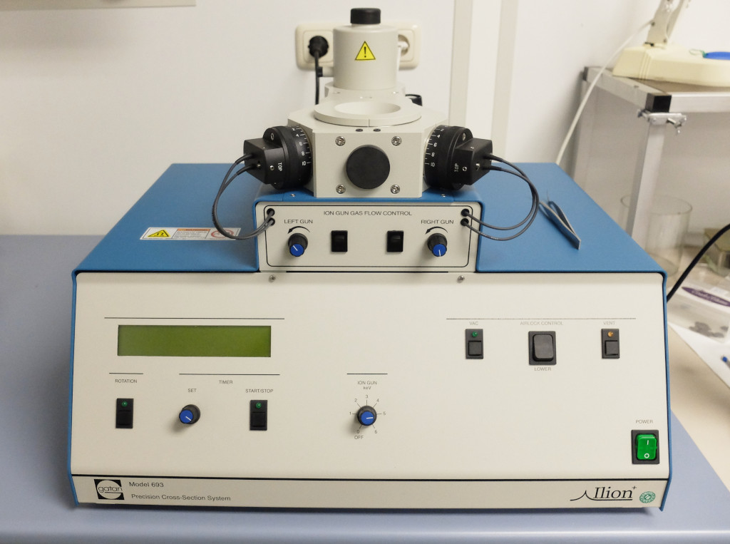
Gatan Ilion cross-section polishing for scanning electron microscopy


Software#
Mapping material properties in analytical electron microscopy produces large amounts of data, as a spectrum or image is recorded at each pixel. In our case, these are cathodoluminescence spectra and transients (1-2 spatial + 1 signal dimension), but also combined spectral and time-resolved data sets (1-2 spatial + 2 signal dimensions). The efficient handling, targeted evaluation and appropriate visualization of these multidimensional (hyperspectral) data sets requires suitable tools.
The PDI is involved in the development of the open-source Python package HyperSpy and some of its extensions. In particular, we initiated the extension package LumiSpy together with the CL group at the University of Cambridge in 2020, which we continue to develop further - focusing on time-resolved luminescence spectroscopy within the framework of ZALKAL. These Python packages are jointly programmed by an international team of scientists and used by many colleagues worldwide. On this basis, even complex analysis routines can be programmed and reliably reproduced with little effort.
The LumiSpy extension supplements HyperSpy with specific functions for cathodoluminescence and photoluminescence data, but is also useful for data from other methods, e.g. Raman spectroscopy, electroluminescence and Fourier transform infrared spectroscopy. This includes e.g. the conversion of the data axes units.
RosettaSciIO enables the reading and saving of scientific data formats from electron microscopy and beyond. Both overarching formats and many manufacturer-specific file types are supported. Important metadata is also read in and made available in a standardized tree structure.